

| Technical Specifications | ||
|---|---|---|
| Product Name | Gel Imaging System | |
| Cat. No. | BE-SCG-W1000 PLUS | |
| Dimensions | 400 × 371 × 700mm | |
| Camera | Pixel Resolution | 6.3 million pixels |
| Resolution | 3072 × 2048 | |
| Pixel Size | 2.4 × 2.4μm | |
| Target Size | 1/1.8” (7.37 × 4.92mm) | |
| Full Well Capacity | 10.4ke- | |
| Sensitivity | 760mv | |
| Readout Noise | 2.14e- | |
| Dark Current | 0.15mV | |
| Signal-to-Noise Ratio | 40.2dB | |
| Exposure Time | 17us-15s | |
| Binning Mode | 1 × 1, 2 × 2, 3 × 3, 4 × 4 | |
| Grayscale | Selectable 8-bit (256 grayscale levels) or 16-bit (65,535 grayscale levels) settings. | |
| Camera Type | Color Camera | |
| Lens | Aperture | F1.0-F16 |
| Focal Length | 8-48mm | |
| Type | Motorized Zoom Lens | |
| Close-up lens | 2 times | |
| Filter | 590/60nm | |
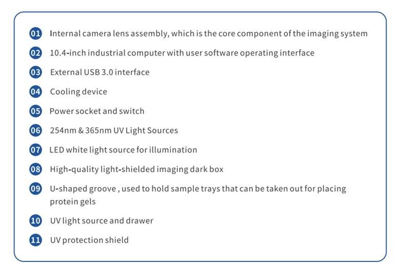
| Light Source | Bright Field Light Source | Downward-facing LED white light source, symmetrically distributed on both sides. |
| UV Light Source | 310 nm LED array, uniform transmission illumination. Epi-illumination 254 nm / 365 nm ultraviolet LED light source, symmetrically distributed on both sides. | |
| Blue/White Dual Light Sources | Blue/white transmission switching, each with 3-level cyclically adjustable power. | |
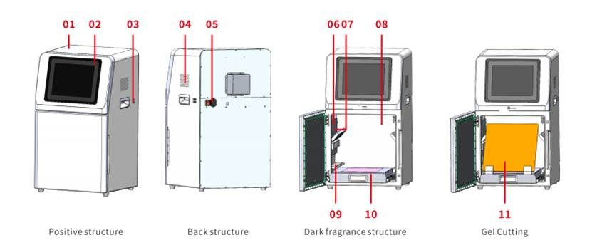
| Dark Box | Brightfield Light Source | Epi-illumination LED white light source, symmetrically distributed on both sides. |
| Door Control | Door control sensor can control the on/off of the bright field light source. | |
| Field of View | Effective field of view is 240 × 240 mm. | |
| Gel Cutting | After opening the door, the UV light source can be pulled out for cutting, in conjunction with a UV protection shield. | |
| Software Functions | Camera Settings | Adjustable contrast and exposure time. |
| Lens Adjustment | Choose adjustment range: coarse adjustment, fine adjustment, super-fine adjustment. Lens: Zoom, focus, aperture adjustment. | |
| Mode Selection | Nucleic Acid Gel Mode, Protein Gel Mode, Gel Cutting Mode. | |
| Image Cropping | Image cropping: Crop the necessary parts of the bands. | |
| Industrial Computer | 10.4"display(1024x768) Windows 100S 16GB RAM, 512GBSSD, Integrated Bluetooth/Wi-Fi. |
|
| External Interfaces | USB 3.0×2 | |
| Operating Voltage | 90-132VAC/180-264VAC (selected via switch).47-63 Hz | |
| Product Power | ≤300W | |
| Product Net Weight | 30.65Kg | |
| Cat. No. | BE-SCG-W5000 PLUS | BE-SCG-W3000 PLUS | BE-SCG-W1000 PLUS |
|---|---|---|---|
| Dimension | 400 × 371 × 700 mm | 400 × 371 × 700 mm | 400 × 371 × 700 mm |
| Camera | Depth-cooled high sensitivity camera | Depth-cooled high sensitivity camera | High-Sensitivity Camera |
| Resolution | 2992 × 3000, 9 megapixels | 2992 × 3000, 9 megapixels +2992 × 3000 45 megapixels |
3072 × 2048, 6.3 megapixels |
| Pixel | 3.76 × 3.76 μm | 3.76 × 3.76 μm | 2.4 × 2.4 μm |
| Shooting Area | Effective field of view for blotting film/protein gel: 136 × 136 mm Effective field of view for nucleic acid gel: 140 × 140 mm. |
Blotting Film 136 × 136 mm | Nucleic Acid Gel / Protein Gel 140 × 140 mm |
| Cooling Temperature | Relative ambient temperature -40°C | Relative ambient temperature -40°C | - |
| Light Source | Bright-field Light Source: Downward-facing LED white light source, symmetrically distributed on both sides. UV Light Source: 310 nm LED array for uniform transmission illumination. |
Downward-facing LED white light, symmetrically distributed on both sides. | Bright-field Light Source: Downward-facing LED white light source, symmetrically distributed on both sides. UV Light Source: 310 nm LED array for uniform transmission illumination. |
| Industrial Computer | 1 match 0.4 inches, 1024×768 Windows operating system |
10.4 inches, 1024 × 768 Windows operating system |
10.4 inches, 1024 × 768 Windows operating system |
| External Interface | 2 USB3.0 | 2 USB3.0 | 2 USB3.0 |
| Working Voltage | 90~132VAC/180~264VAC (Selectable via switch )47~63HZ | 90-132V/180-264V | 90~132VAC/180~264VAC (Selectable via switch) 47~63HZ |
| Product Power | 200 W | ≤200 W | 200 W |
| Net Weight | 25 kg | 23.45 kg | 30 kg |
| Real-Time Imaging | Yes | Yes | - |
| Time Imaging | Yes | Yes | - |
| Time Accumulation | Yes | Yes | - |
| Auto Exposure | Yes | Yes | Yes |
| Choice of 3 Imaging Modes | Yes | Yes | - |
| Protein Gel/Nucleic Acid Gel Imaging | Yes | - | Yes |
| Nucleic Acid Gel Cutting | Yes | - | Yes |

| Product Name | Chemiluminescence Imaging System | |
|---|---|---|
| Cat. No. | SCG-W3000 PLUS | |
| Dimensions | 400mm × 371mm × 700mm | |
| Camera 1 | Pixel Resolution | 9 million pixels |
| Resolution | 2992*3000 | |
| Pixel Size | 3.76μm×3.76μm | |
| Target Size | 1“ (11.28mm×11.28mm) | |
| Full Well Capacity | 16.5ke-(HCG), 50.5ke-(LCG) | |
| Sensitivity | 877mv@1/30s | |
| Readout Noise | 1.24e-(HCG), 3.22e-(LCG) | |
| Dark Current | 0.0003e-/s/pixel@-15℃ | |
| Signal-to-Noise Ratio | 42.2dB (HCG), 47dB (LCG) | |
| Exposure Time | 0.1ms~1h | |
| Binning Mode | 1×1, 2×2, 3×3 | |
| Grayscale | 16-bit (65536 levels) | |
| Cooling | Relative to Ambient Temperature-40℃ | |
| Camera Type | Black and White Camera | |
| Camera 2 | Pixel Resolution | 45 million pixels |
| Resolution | 2992*3000 | |
| Pixel Size | 2.315×2.315μm | |
| Target Size | 1.4“ (18.93×13 mm) | |
| Full Well Capacity | 10.8ke- | |
| Sensitivity | 419mv | |
| Readout Noise | 2.12e- | |
| Dark Current | 0.12mV | |
| Signal-to-Noise Ratio | 40.3dB | |
| Exposure Time | 17μs~15s | |
| Binning Mode | 1×1, 2×2, 3×3 | |
| Grayscale | 8bit(256 gray) | |
| Camera Type | Color Camera | |
| Lens 1 | Aperture | F0.95-F16 |
| Focal Length | 17mm | |
| Type | Prime lens | |
| Light Source | Bright Field Light Source | Downward-facing LED white light source, symmetrically distributed on both sides. |
| Dark Box | Light Isolation | Fully light-sealed, isolates environmental light. |
| Door Control | Door control sensor can control the on/off of the bright field light source. | |
| Field of View | Effective field of view is 136mm*136mm (expandable to 200mm×200mm if needed). | |
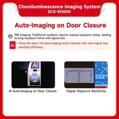
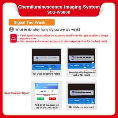
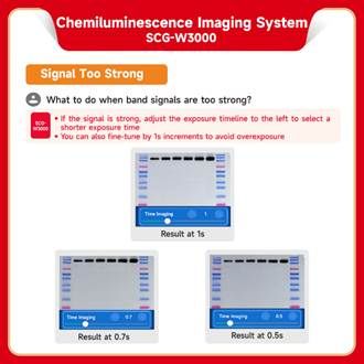
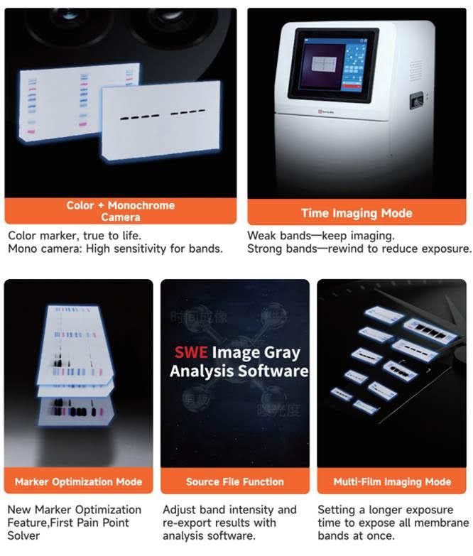
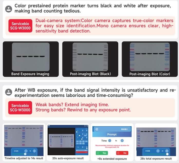
| Auto Exposure | Intelligent exposure technology quickly determines the optimal exposure time and automatically performs binning. Combined with time-lapse imaging and time-accumulation functions, users can obtain the best image results with just one operation. | |
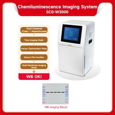
| Software Functions | Exposure Modes | High Quality: Highest image quality. Standard: Balances image quality and exposure speed. High Sensitivity: Fastest exposure speed. |
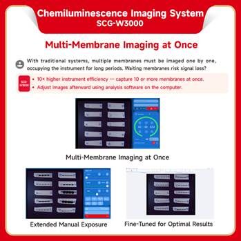
| Real-Time lmaging | Real-time display of changes in sample signals during the exposure process allows users to observe every detail of the capture. Areas of overexposure are clearly indicated when samples exceed the optimal exposure range. | |
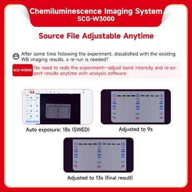
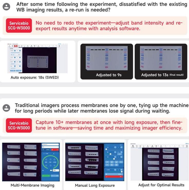
| Time Imaging | After the exposure is complete, every frame image captured during the exposure period can be generated. With precise retrospective adjustments, users can select any frame within that exposure time as the final output. | |
| Time Accumulation | For samples with insufficient exposure, users can continue the exposure after the initial run is completed, allowing the sample to receive additional exposure time on top of the original exposure. | |
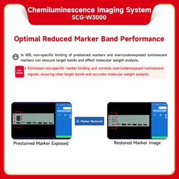
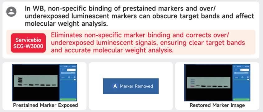
| Optional Settings | Color Marker/Black & White Marker, Overexposure Prompt/No Overexposure Prompt |
|
| Industrial Computer | 10.4-inch display with a resolution of 1024 x 768, running on Windows 10 operating system, featuring 16GBof RAM, 512 GBssD, built-in Bluetooth, and WiFi. | |
| External Interfaces | 2 USB 3.0 ports | |
| Operating Voltage | 90~132VAC/180~264VAC (selectable via switch),47~63Hz. | |
| Product Power | ≤200W | |
| Product Net Weight | 23.45Kg | |
| Cat. No. | BE-SCG-W5000 PLUS | BE-SCG-W3000 PLUS | BE-SCG-W1000 PLUS |
|---|---|---|---|
| Dimension | 400 × 371 × 700 mm | 400 × 371 × 700 mm | 400 × 371 × 700 mm |
| Camera | Depth-cooled high sensitivity camera | Depth-cooled high sensitivity camera | High-Sensitivity Camera |
| Resolution | 2992 × 3000, 9 megapixels | 2992 × 3000, 9 megapixels +2992 × 3000 45 megapixels |
3072 × 2048, 6.3 megapixels |
| Pixel | 3.76 × 3.76 μm | 3.76 × 3.76 μm | 2.4 × 2.4 μm |
| Shooting Area | Effective field of view for blotting film/protein gel: 136 × 136 mm Effective field of view for nucleic acid gel: 140 × 140 mm. |
Blotting Film 136 × 136 mm | Nucleic Acid Gel / Protein Gel 140 × 140 mm |
| Cooling Temperature | Relative ambient temperature -40°C | Relative ambient temperature -40°C | - |
| Light Source | Bright-field Light Source: Downward-facing LED white light source, symmetrically distributed on both sides. UV Light Source: 310 nm LED array for uniform transmission illumination. |
Downward-facing LED white light, symmetrically distributed on both sides. | Bright-field Light Source: Downward-facing LED white light source, symmetrically distributed on both sides. UV Light Source: 310 nm LED array for uniform transmission illumination. |
| Industrial Computer | 1 match 0.4 inches, 1024×768 Windows operating system |
10.4 inches, 1024 × 768 Windows operating system |
10.4 inches, 1024 × 768 Windows operating system |
| External Interface | 2 USB3.0 | 2 USB3.0 | 2 USB3.0 |
| Working Voltage | 90~132VAC/180~264VAC (Selectable via switch )47~63HZ | 90-132V/180-264V | 90~132VAC/180~264VAC (Selectable via switch) 47~63HZ |
| Product Power | 200 W | ≤200 W | 200 W |
| Net Weight | 25 kg | 23.45 kg | 30 kg |
| Real-Time Imaging | Yes | Yes | - |
| Time Imaging | Yes | Yes | - |
| Time Accumulation | Yes | Yes | - |
| Auto Exposure | Yes | Yes | Yes |
| Choice of 3 Imaging Modes | Yes | Yes | - |
| Protein Gel/Nucleic Acid Gel Imaging | Yes | - | Yes |
| Nucleic Acid Gel Cutting | Yes | - | Yes |


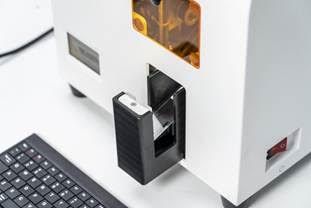
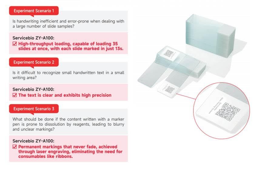
| Product Model | BE-ZY-A100 |
|---|---|
| Module Specifications | Slides |
| Laser Wavelength | 355nm Ultraviolet Laser |
| Laser Power and Heat Dissipation |
3Wair-cooled ultraviolet solid-state laser, air-cooled |
| Laser Average Power | >3W@40kHz |
| Dimensions | 540×315×545mm |
| Weight | 25 kg |
| Operating Function | Prints the imported information onto the product using a laser. |
|---|---|
| Marking Working Temperature | Temperature: 15-35°Humidity <90% |
| Display Screen | 10,1"1280 x 800@60 Hz, LED backlight, capacitive touchscreen. |
| User-Friendly Display of Operating Status | Yes, with a dynamic temperature color background, animated operating status. |
| Interface | Minimalist Interface. |
| Ethernet Port | Built-in Gigabit Ethernet Port |
| Intelligent Self-Check on Startup | Yes, the system interface can also start self-check. |
| Marking Information Recording | Yes |
| Fault Self-Diagnosis and Reporting | Yes |
| Instrument Maintenance Index Function | Yes, evaluates product maintenance based on instrument usage conditions. |
| System Time | Yes, can set instrument date and time. |
| Screen Brightness Adjustment | Yes, adjustable in multiple levels. |
| Language Selection | Yes, switch system language (Chinese/English). |
| System Interface | Includes system settings, self-check, restore factory settings, software upgrade, engineering interface (limited to factory mode), etc. |
| Help System | Product features, software characteristics, quick guide, fault codes, and contact information. |
| WeChat Service Platform | Yes, when a fault occurs, users can send fault photos to the company's WeChat service platform via mobile phone. |
| Factory Mode | Yes, it is accessible only to manufacturers with special passwords. It allows system data viewing, settings adjustment, parameter calibration, and configuration. |